

Researchers of the University College of London reported that the invention detects disorders in the musculoskeletal system in a few minutes.
European physicists have created a device that makes it possible to obtain three-dimensional photoacoustic images of human internal organs using portable laser scanners, which can be used to assess the state of various human body tissues in real time. This was reported by the press service of the University College London (UCL).
"Our main breakthrough is that we managed to speed up image acquisition by about 100-1000 times compared to the speed at which other photoacoustic tomography systems work. This makes it possible to avoid smearing associated with movement and obtain the most detailed images in real time, which paves the way for the visualization of physiological processes, – explained UCL professor Paul Bird, whose words are cited by the university's press service.
For many years, researchers have been working on the creation of so-called photoacoustic tomography systems. They make it possible to obtain images of the internal organs of the body with the help of sets of lasers that produce beams of ultrasonic waves during interaction with hemoglobin in blood cells. These oscillations are captured and processed by several highly sensitive ultrasound sensors and combined into a three-dimensional or two-dimensional image.
200% Deposit Bonus up to €3,000 180% First Deposit Bonus up to $20,000Such systems allow obtaining very detailed images of internal organs with a resolution of several micrometers, but previously for their work the subject or patient was required to remain motionless for the very long time required to fully reconstruct a three-dimensional image of their body tissues. European physicists discovered that this process can be accelerated by several orders of magnitude with the help of an optical detector of ultrasonic vibrations created by them.
It is a set of two special mirrors and a flexible polymer layer separating them, which captures vibrations generated by ultrasound in the thickness of the patient's body and transmits them to the mirrors. The shape of the mirrors changes under the influence of these vibrations, which makes it possible to capture them using a set of many laser beams that irradiate the mirrors. This makes it possible to obtain three-dimensional images of organs in a few seconds and obtain two-dimensional images and videos in real time.
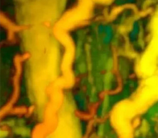
Scientists have successfully tested the operation of this scanner in experiments on the condition of the vessels of the limbs, deep layers of the skin, as well as the joints of the hands and fingers of 10 patients who suffered from diabetes, rheumatoid arthritis and breast cancer. In this way, it is possible to detect the presence of foci of inflammation, arthritis and other disorders in the work of the musculoskeletal system in a few minutes, while previously such a diagnosis took about an hour, which significantly increases the comfort of this procedure for patients, the researchers concluded.